Your Oral Cancer Histopathology Report, Explained

Need expert consultation? Book an appointment with Dr. Pradeep S. or Dr. Kalpa Pandya.
Book AppointmentWaiting for the final pathology result after oral cancer surgery is one of the most anxious parts of treatment. Your oral cancer histopathology report is the document that answers the most important question after the operation: was surgery enough, or is radiation or chemoradiation needed? This guide explains each section of the report in plain language so you can follow the conversation with your surgeon.
This is the report on the tissue removed at surgery. If you are still at the diagnosis stage, our oral cancer biopsy guide explains the smaller diagnostic biopsy instead.
Biopsy Report vs. Final Histopathology Report
The first biopsy was a small sample taken to confirm that cancer is present and identify its type. It is effectively a "yes or no" test that starts treatment planning.
The final histopathology report is produced after surgery, when the pathologist examines the whole tumour, the rim of healthy tissue around it and, if a neck dissection was done, the lymph nodes. A biopsy is like looking through a keyhole; the final report is like walking into the room. It shows the tumour's true size, how deep it grew, whether it reached nerves or vessels, and how close it came to the cut edges. Because it decides whether you need treatment after surgery, it is the most important document in your post-operative file.
Frozen Section vs. the Final Report
During the operation, your surgeon may send small pieces of tissue from the edges of the resection for a frozen section. The tissue is rapidly frozen, cut and examined while you are still under anaesthesia, usually within about half an hour. If cancer is seen at an edge, the surgeon can remove more tissue in the same operation.
Frozen section is a useful real-time check, but it has limits. Freezing distorts the tissue slightly, only selected areas are sampled, and the pathologist has minutes rather than days. The final report is made on tissue that has been fixed in formalin, processed into wax blocks and stained, with the whole specimen examined. Occasionally the final report finds something the frozen section did not, or the reverse. When the two differ, the final report is the one your treatment plan is based on.
Tumour Type and Grade
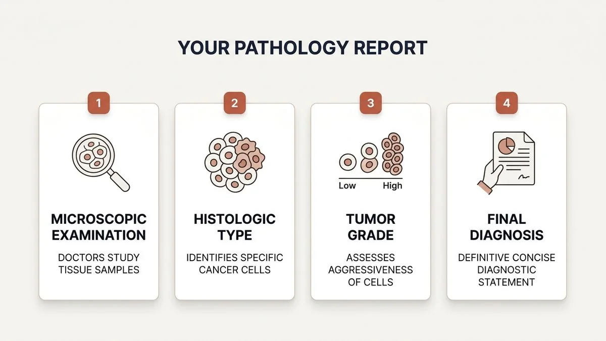
The report begins with the tumour type. In the mouth, the large majority of cancers are squamous cell carcinoma (SCC), arising from the lining cells.
It then gives a grade, which describes how closely the cancer cells resemble normal cells:
- Well differentiated: cells look fairly similar to normal lining cells and tend to grow more slowly.
- Moderately differentiated: cells have lost more of their normal appearance.
- Poorly differentiated: cells look very abnormal and disorganised and may behave more aggressively.
Worst Pattern of Invasion (WPOI)
Many pathologists now also report the worst pattern of invasion, which describes how the tumour's leading edge invades. A broad, pushing front is more favourable; small scattered islands or single cells at the edge suggest more aggressive behaviour and a higher chance of spread or recurrence. For an example of how site and staging fit together, see our guide to tongue SCC biopsy and staging.
Tumour Size and Depth of Invasion (DOI)
Depth of invasion is one of the most important numbers in the report, and it is often confused with tumour thickness.
- Tumour thickness is measured from the surface of the tumour to its deepest point.
- Depth of invasion is measured from the level of the basement membrane of the nearby normal lining down to the deepest point of the tumour.
Under the AJCC 8th edition staging system, DOI changes the T category: tumours deeper than 5 mm and deeper than 10 mm are placed in higher T categories even if they look small on the surface. Deeper tumours carry a higher risk of hidden spread to the neck, which is one reason the neck is so often treated in oral cancer. Our article on when mouth cancer needs a neck dissection explains that decision.
Surgical Margins: Clear, Close and Positive
Margins describe the healthy tissue removed around the tumour. During a wide local excision the aim is a clear rim of normal tissue on every side. The report measures the distance from the cancer to the nearest cut edge:
- Clear: generally more than 5 mm. This is the target and carries a low risk of the cancer returning at that site.
- Close: roughly 1 to 5 mm. The cancer came near the edge, and postoperative radiotherapy is often discussed.
- Positive (involved): tumour cells reach the inked cut edge. Some centres also treat a margin under 1 mm as involved. A positive margin almost always means further treatment.
Margins are reported for each direction (front, back, deep and so on), so ask which margin was closest and by how much.
Perineural and Lymphovascular Invasion
Beyond size and margins, the pathologist looks at how the cancer has interacted with nerves and vessels.
- Perineural invasion (PNI): cancer cells growing along or around a nerve. Nerves can act as pathways for spread, so PNI signals a higher risk of the cancer returning locally.
- Lymphovascular invasion (LVI): cancer cells inside lymphatic channels or small blood vessels, a route towards the lymph nodes and beyond.
Finding PNI or LVI often tips the balance towards radiotherapy after surgery, even for a smaller tumour.
Have your report but not sure what it means? Dr. Pradeep S. and Dr. Kalpa Pandya review oral cancer pathology reports at Apollo Main Hospital, Greams Road, Chennai. Book a consultation or call +91 96633 03747 to go through your results and next steps.
Lymph Nodes and Extranodal Extension (ENE)
If you had a neck dissection, the report lists how many lymph nodes were removed, how many contained cancer, and the size of the largest deposit. Lymph node status is one of the strongest predictors of outcome in oral cancer.
The report will also state whether there is extranodal extension (ENE): cancer that has broken through the capsule of a lymph node into the surrounding tissue. Pathological ENE raises the N category and is a strong signal that chemotherapy should be added to radiation after surgery.
Putting It Together: pTNM Staging
Before surgery you were given a clinical stage (cTNM) based on examination and scans. The histopathology report allows a final pathological stage (pTNM), where "p" stands for pathological. It combines:
- pT: tumour size and depth of invasion
- pN: number, size and features of involved lymph nodes, including ENE
- M: spread to distant organs, if known
Why the Clinical and Pathological Stage Can Differ
It is common for the stage to change after surgery, in either direction, because the specimen gives far more detail than any scan:
- Depth of invasion: scans and examination estimate depth, but only the microscope measures it to the millimetre, so a tumour may move up or down a T category.
- Hidden nodal disease: lymph nodes can contain small deposits of cancer that look completely normal on CT, MRI or PET-CT. Finding these raises the N category.
- Reactive nodes: the opposite also happens. A node that looked enlarged on a scan may turn out to be inflamed rather than cancerous, lowering the N category.
- ENE: extranodal extension can be suspected on imaging but is only confirmed under the microscope.
A change in stage does not mean the earlier assessment was wrong. It means the team now has the most accurate information available. Our guide to oral cancer stages explains the stage groups, and our article on stage 1 mouth cancer covers the early end of the scale. The final pathological stage is discussed at the multidisciplinary tumour board, where surgeons, radiation oncologists and medical oncologists agree the next step.
A Worked Example: Reading a Report Line by Line
The example below is illustrative only. It is not a real patient's report, and real reports vary in layout and wording. It shows how each line translates into plain language.
Sample summary: "Right lateral tongue, partial glossectomy with neck dissection. Squamous cell carcinoma, moderately differentiated. Tumour size 2.6 cm; DOI 7 mm. Closest margin 3 mm (deep); all other margins more than 5 mm. PNI present; LVI absent. 2/34 lymph nodes positive (level II and III, right), ENE absent. pT2 pN2b."
- Squamous cell carcinoma: the usual type of mouth cancer, arising from the lining cells.
- Moderately differentiated: the cells look moderately abnormal, which sits in the middle of the grading scale.
- Tumour size 2.6 cm, DOI 7 mm: the tumour is between 2 and 4 cm across and has grown 7 mm below the normal lining. Under AJCC 8th edition rules, this combination is T2.
- Closest margin 3 mm (deep): every edge is clear of cancer, but the deep edge is close (1 to 5 mm). The other edges are clear.
- PNI present: cancer cells were seen around a nerve, a feature linked to a higher risk of local recurrence.
- LVI absent: no cancer cells were seen inside lymphatic or blood vessels.
- 2/34 nodes positive, ENE absent: 34 lymph nodes were examined and 2 contained cancer, both on the same side as the tumour. Neither had broken out of its capsule.
- pT2 pN2b: the final pathological stage. pN2b means cancer in more than one lymph node on the same side, none larger than 6 cm, without ENE. With no distant spread, this corresponds to stage IVA.
What it usually means: a close margin, PNI and positive lymph nodes are each reasons for postoperative radiotherapy. Because the margins are not involved and there is no ENE, chemotherapy would not usually be added to the radiation. Your own team would confirm this after reviewing the full report.
What Your Report Means for Radiation or Chemoradiation
The main purpose of the report is to decide whether surgery alone is enough. Based on NCCN guidelines for oral cavity cancer, each finding usually maps to a next step as follows:
- Positive (involved) margin: chemoradiation is usually recommended; re-excision is considered if it is feasible.
- Extranodal extension (ENE): chemoradiation is usually recommended.
- Close margin: postoperative radiotherapy is usually considered.
- Perineural invasion (PNI): postoperative radiotherapy is usually considered.
- Lymphovascular invasion (LVI): postoperative radiotherapy is usually considered.
- pT3 or pT4 tumour: postoperative radiotherapy is usually recommended.
- pN2 or pN3 nodal disease: postoperative radiotherapy is usually recommended.
- A single small positive node (pN1) without other adverse features: postoperative radiotherapy is considered case by case.
- Small tumour (pT1 or pT2), clear margins, node-negative, no adverse features: surgery alone with close follow-up is often enough.
Timing matters: postoperative radiotherapy should ideally begin within about 6 weeks of surgery, so your recovery and the radiation plan are coordinated. These are guidelines, not rules; the final recommendation also considers your general health and recovery. If the cancer returns later, our guide to options for advanced and recurrent mouth cancer explains what happens next.
Questions to Ask About Your Report
Bring the report to your post-operative review and ask:
- What were my closest margins, in millimetres, and in which direction?
- What was the depth of invasion?
- Was there perineural or lymphovascular invasion?
- How many lymph nodes were removed, how many were involved, and was there ENE?
- Did my stage change from the clinical stage before surgery?
- Based on this report, do I need radiotherapy or chemoradiation, and when would it start?
Getting Your Report Reviewed in Chennai
A histopathology report is technical, and it is reasonable to want it explained carefully or reviewed again. Dr. Pradeep S., an oral and maxillofacial surgeon with fellowship training in head and neck surgical oncology and a postgraduate diploma in clinical research, and Dr. Kalpa Pandya go through these reports with patients at Apollo Main Hospital, Greams Road.
If your surgery was done elsewhere, our second opinion service explains how to share records, including from outside Chennai.
How a Second Pathology Review Works
A second review means a pathologist looks again at the original tissue, not just at the written report. To make that possible, ask the hospital where you had surgery for:
- The histopathology report: the final signed report, plus the earlier biopsy report.
- The stained slides: the glass slides the first pathologist examined.
- The paraffin blocks: the wax blocks the slides were cut from. Hospitals normally release these on written request, sometimes for a deposit, because they are kept as part of your record.
- The operation notes: these describe what was removed and how the specimen was oriented.
- Your scans: CT, MRI or PET-CT reports and images from before surgery.
With the blocks, the reviewing pathologist can cut fresh sections or run additional stains if a feature such as perineural invasion or ENE is uncertain. The review usually confirms the original findings, but sometimes it refines a margin measurement or a node count in a way that changes the recommendation for radiation. Specific guidance by site is also available for tongue cancer treatment and cheek cancer treatment.
Glossary of Pathology Terms
- Squamous cell carcinoma (SCC): the most common oral cancer, arising from the lining cells of the mouth.
- Dysplasia: pre-cancerous changes in the lining that are not yet invasive cancer.
- Grade (differentiation): how closely cancer cells resemble normal cells.
- Depth of invasion (DOI): how deep the tumour has grown below the normal lining; it affects the T stage.
- Margin: the distance between the cancer and the cut edge of the removed tissue.
- Perineural invasion (PNI): cancer growing along a nerve.
- Lymphovascular invasion (LVI): cancer cells inside lymphatic or blood vessels.
- Extranodal extension (ENE): cancer breaking out of a lymph node's capsule.
- pTNM: the final pathological stage after surgery.
- Adjuvant therapy: radiation or chemotherapy given after surgery to lower the risk of recurrence.
References
- Amin, M. B., et al. AJCC Cancer Staging Manual, 8th edition. Springer, 2017.
- National Comprehensive Cancer Network (NCCN). "NCCN Guidelines for Patients: Oral Cancers." https://www.nccn.org/patients
- D'Cruz, A. K., et al. "Elective versus Therapeutic Neck Dissection in Node-Negative Oral Cancer." New England Journal of Medicine, 2015.
If you would like your report explained, book an appointment or call +91 96633 03747.
Next step
Concerned about a symptom you read here? Get a specialist opinion in Chennai.
Dr. Pradeep S. and Dr. Kalpa Pandya consult at Apollo Hospitals, Greams Road. Same-day responses on WhatsApp for most enquiries.

Dr. Pradeep S.
MDS (OMFS) · FHNS · FIBCSOMS

Dr. Kalpa Pandya
MDS (OMFS) · FHNS — Head & Neck Oncology
Authored by
Medically reviewed by