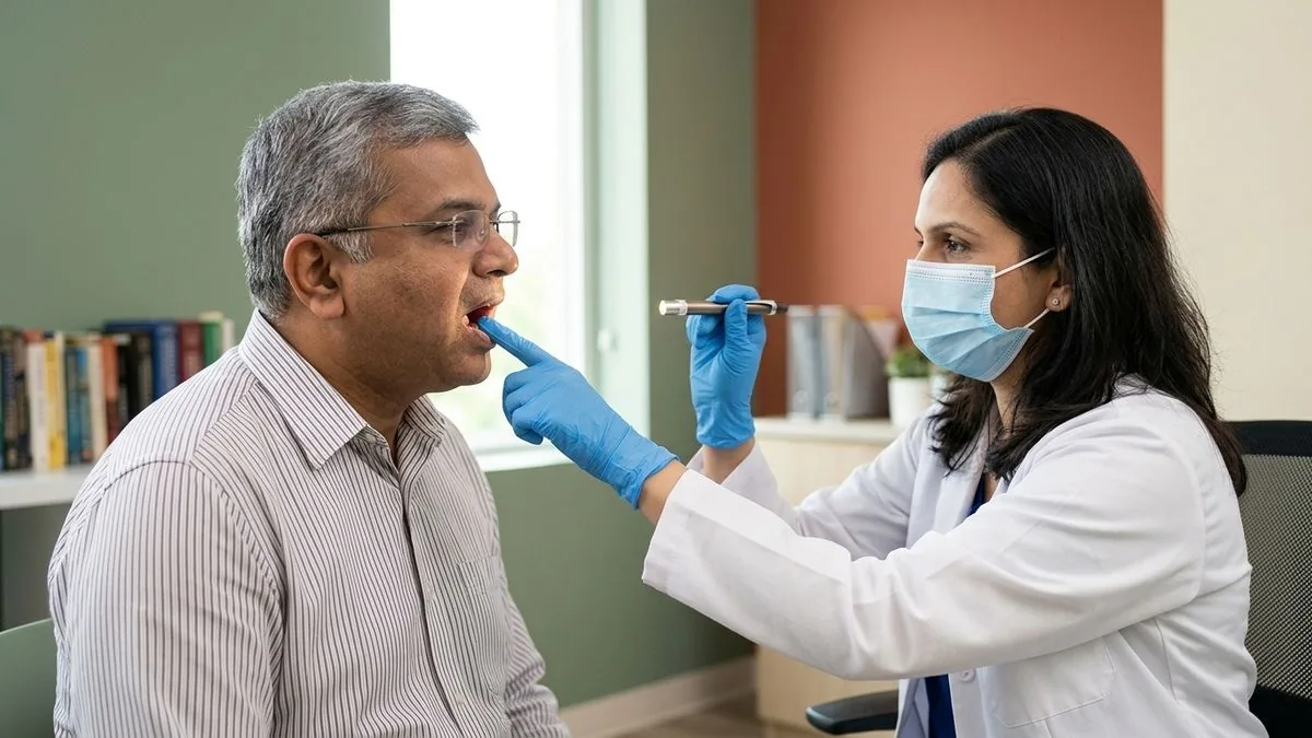
Buccal mucosa cancer (inner cheek) is strongly linked to tobacco and areca-nut use and is common in India. Wide excision with margins, neck dissection where needed, and reconstruction restore form and function.
Buccal Mucosa Cancer Treatment: Cancer Inside the Cheek
Buccal mucosa cancer — cheek cancer, or mouth cancer inside the cheek — starts in the soft lining on the inner surface of the cheeks. Buccal mucosa cancer treatment is mainly surgical: the tumour is removed with a safe margin of healthy tissue, the neck nodes are treated when needed, and the cheek (and jaw, if involved) is rebuilt so that you can open your mouth, chew and speak.
In India this is one of the commonest cancers surgeons see. It is closely tied to chewing tobacco, gutka, paan and areca nut, and it often develops on a background of long-standing white patches or oral submucous fibrosis. Early stage cheek cancer can be cured in most patients; the difficulty is that it is often painless at first and is noticed late.
This page explains how cheek cancer is recognised, why it is so common here, how it is staged, the operations involved, reconstruction choices, when radiotherapy is added, survival figures from Indian data, and what recovery involves.
Early Signs of Cheek Cancer vs an Ordinary Ulcer
An ordinary ulcer from biting the cheek or rubbing on a sharp tooth usually heals within one to two weeks. Cheek cancer does not. See a specialist if any of these persist beyond two weeks:
A non-healing ulcer: A sore on the inner cheek that keeps returning or slowly grows, often with raised, firm edges.
A rough or thickened area: A patch that feels hard or warty to the tongue, or a lump within the cheek.
White or red patches:Leukoplakia (white) or erythroplakia (red) that change in size, colour or texture.
Restricted mouth opening (trismus): Gradually opening less wide than before. It can mean the tumour has reached the chewing muscles, or that oral submucous fibrosis is present.
Pain or bleeding: Burning with spicy food, bleeding on touch, or loosening of teeth near the patch, which can mean the gum or jawbone is involved.
A lump in the neck: A firm swelling under the jaw may be the first sign of spread to the lymph nodes.
Worldwide, the tongue is the commonest site of mouth cancer. In India it is the buccal mucosa and the neighbouring gum — together called the gingivobuccal complex. The reason is where people hold tobacco: a quid of betel leaf, areca nut, khaini or gutka is kept against the inner cheek for hours, exposing the lining to carcinogens day after day.
The scale is large. India accounts for about a third of the world's oral cancers, and according to the Global Adult Tobacco Survey (GATS-2, 2016–17), 21.4% of Indian adults use smokeless tobacco. Our guide to gutka and pan masala risk explains how these products damage the mouth.
Many patients also have oral submucous fibrosis (OSMF), a stiffening of the cheek lining caused by areca nut. OSMF restricts mouth opening and is itself a pre-cancerous condition. Stopping all tobacco and areca nut — before and after treatment — is one of the most important things you can do to reduce the risk of a new cancer.
Pre-Cancer in the Cheek: White Patches, Red Patches and OSMF
Most cheek cancers do not appear out of nowhere. They are often preceded for months or years by changes in the lining that doctors call oral potentially malignant disorders. Recognising and treating these is the best chance of preventing cancer altogether.
Leukoplakia: A white patch that cannot be wiped off and has no other obvious cause. Most never become cancer, but pooled studies suggest roughly 6–7% eventually do, and the risk is higher for patches that are thick, speckled or growing.
Erythroplakia: A red, velvety patch. It is less common than leukoplakia but far more likely to contain severe dysplasia or early cancer, so it should always be biopsied.
Oral submucous fibrosis: Progressive stiffness and pale, leathery bands in the cheek from areca nut use. Studies following patients over years report malignant change in roughly 4–6%.
A biopsy shows whether a patch has dysplasia (abnormal but not yet cancerous cells) and how severe it is. Mild changes may be watched closely after stopping tobacco; more severe changes or suspicious areas are usually removed. If a patch has already turned into cancer, finding it at this stage often means a smaller operation and a much better outlook.
How Buccal Mucosa Cancer Is Diagnosed and Staged
Diagnosis begins with an examination of the cheek, gums, jaw and neck, checking how deep the lump extends, whether it is fixed to bone, and how wide the mouth opens. A biopsy taken under local anaesthetic confirms the diagnosis and grade.
Scans then map the extent. A contrast CT (CECT) is particularly good at showing whether the jawbone is eroded; MRI shows spread into soft tissues and the chewing muscles. Larger tumours may need a chest scan or PET-CT. Staging follows the AJCC 8th edition TNM system: the tumour's size and depth of invasion, lymph node spread and any distant spread combine into stages I to IV.
Bone involvement is the key surgical question in cheek cancer. Tumours that sit close to the gum often reach the jaw, and that decides whether a rim of bone or a full segment needs to be removed.
Buccal Mucosa Cancer Surgery: Excision, Jaw Surgery and Neck Dissection
Surgery is the main treatment. The aim is complete removal with clear margins in every direction — through the cheek, towards the skin, and down to bone — while preserving mouth opening and appearance as far as possible.
Wide local excision: The tumour is removed with a cuff of normal tissue, with margins checked by frozen section during the operation. See wide local excision.
Marginal mandibulectomy: When the tumour touches but has not deeply invaded the jaw, a rim of bone is removed while keeping the lower jaw continuous.
Segmental mandibulectomy: When the cancer has grown into the bone, a full section of the jaw is removed and must be rebuilt. Our mandibulectomy page explains both types.
Composite resection: For advanced tumours, cheek, jaw and neck tissues are removed together in one block — sometimes called a commando operation. See composite resection.
A neck dissection is usually performed at the same operation. Cheek cancers can spread to the neck lymph nodes before they can be felt, and a Tata Memorial Centre trial showed that treating the neck early in node-negative oral cancer improves survival.
Reconstruction: PMMC Flap vs Free Flap
Removing a cheek cancer leaves a gap in the lining of the mouth, sometimes extending through to the skin or including part of the jaw. Rebuilding it stops the mouth scarring shut and restores chewing, speech and facial contour.
PMMC flap: The PMMC flap (pectoralis major myocutaneous flap) uses chest muscle and skin, swung up to the cheek on its own blood supply. It is reliable and robust and does not need microvascular surgery, which makes it valuable for medically frail patients and some salvage situations.
Free flap: In free flap reconstruction, thin tissue from the forearm or thigh — or bone from the leg (fibula) when the jaw has been removed — is transferred and connected to neck vessels under a microscope. This is performed with our reconstructive colleague and usually gives a thinner, more flexible lining and a better jaw shape.
Which option suits you depends on the size of the defect, whether bone is missing, your general health and any previous treatment. Our comparison of free flap vs PMMC goes through the trade-offs.
When Radiotherapy Is Added After Surgery
The final histopathology report on the removed tissue guides what happens next. Our explanation of the oral cancer histopathology report covers each term.
Clear margins and no adverse features: Often no further treatment beyond close follow-up — for example a small tumour with margins over 5 mm and no nodal spread.
Close margins or other risk features: Postoperative radiotherapy is usually recommended for margins of 1–5 mm, nerve or vessel invasion, pT3–T4 tumours or involved nodes.
Positive margins or extranodal extension: Radiotherapy combined with chemotherapy (chemoradiation). A margin under 1 mm is treated as positive.
Radiotherapy should ideally start within about six weeks of surgery. Because the cheek lies close to the teeth and jaw, a dental assessment beforehand is important to reduce the risk of jaw-bone damage later — see dental treatment before radiotherapy.
Buccal Mucosa Cancer Survival by Stage
Indian data is the most useful reference. In Tata Memorial Centre data on 1,895 patients with oral cavity cancer treated in 2016, about 66% of patients with buccal mucosa cancer were alive at five years. Across all oral cavity cancers in that group, five-year survival was about 83% for stage I and about 55% for stage IV.
The biggest influences on outcome are the stage at diagnosis, whether lymph nodes are involved, whether clear margins are achieved, and whether the person stops tobacco. Cheek cancer found as a small ulcer has a very different outlook from one found after the mouth has stopped opening — which is why a persistent patch should never be watched for months.
Cancer of the gum behaves similarly and is often treated alongside cheek cancer; see our guide to gum cancer for that site specifically.
Recovery and Mouth-Opening Exercises
After major surgery with reconstruction, most patients stay in hospital about one to two weeks. A feeding tube supports nutrition while the mouth heals, and a temporary tracheostomy may be used for a few days if swelling is expected.
Keeping the mouth open is the central goal of rehabilitation. Scar tissue and radiotherapy both tend to tighten the cheek, so daily stretching exercises begin as soon as the wound allows — using stacked tongue depressors or a jaw-stretching device — and continue for months. Speech and swallowing therapy, dietary advice and dental rehabilitation follow.
Teeth near the cancer may have to be removed, and losing part of the jaw affects how dentures fit. Once healing is complete, dental rehabilitation — a prosthesis, or in selected patients implants placed in a reconstructed jaw — helps restore chewing. This is planned with your surgical team, because bone that has received radiotherapy needs particular care.
Follow-up is frequent in the first two years, when recurrence is most likely, and continues long term to watch for a new cancer elsewhere in the mouth. Report any new ulcer, lump, pain or reduction in mouth opening between visits rather than waiting for the next appointment.
Questions to Ask Before Cheek Cancer Surgery
A clear conversation before surgery helps you and your family know what to expect. Useful questions include:
Stage and spread: What stage is my cancer, and is the jawbone or are the neck nodes involved?
Extent of surgery: Will part of my jaw be removed, and will both sides of the neck be treated?
Reconstruction: Which flap do you recommend for me, and why?
After surgery: Will I need a feeding tube or tracheostomy, and for how long?
Further treatment: How likely is radiotherapy, and when would it start?
Long term: How will my mouth opening, speech and appearance be affected, and what exercises will I need?
Bringing your biopsy report, scans and a list of your medicines to the first visit makes these answers much more specific.
What Affects the Cost of Buccal Mucosa Cancer Treatment
Cost varies with the extent of treatment. The main factors are:
Size of resection: A wide local excision alone versus a composite resection.
Jaw involvement: Whether a marginal or segmental mandibulectomy is required.
Reconstruction: Direct closure, a PMMC flap or a microvascular free flap, including bone flaps.
Neck dissection and hospital stay: One or both sides of the neck, and the number of days in hospital and intensive care.
Further treatment: Whether radiotherapy or chemoradiation is needed.
We provide a written estimate after reviewing your biopsy and scans at consultation.
Cheek Cancer Surgery in Chennai: What to Expect at Apollo Main Hospital, Greams Road
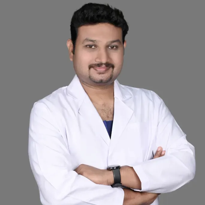
Surgery is led by Dr. Pradeep S., an oral and maxillofacial surgeon with a fellowship in head and neck surgical oncology (FHNS), international board certification (FIBCSOMS) and more than 15 years of experience in oral cancer surgery, including three years in the Department of Head and Neck Oncology at Apollo Proton Cancer Centre. He works with Dr. Kalpa Pandya; both consult at Apollo Main Hospital, Greams Road.
Each plan is agreed with radiation and medical oncologists, pathologists, anaesthetists and rehabilitation teams, so that surgery, reconstruction and radiotherapy follow one another without avoidable delay. Outstation patients can send reports and scans for a remote second opinion before travelling.
Cancer of the tongue is covered separately on our tongue cancer treatment page. To discuss cheek cancer, call +91 96633 03747 or book a consultation for buccal mucosa cancer treatment in Chennai.
Finding us in Chennai
Dr. Pradeep S. and Dr. Kalpa Pandya consult at Apollo Main Hospital, Greams Road, Chennai, where they treat cheek cancer for patients from across Tamil Nadu, West Bengal, Bangladesh and the wider region. Remote second opinions help outstation patients plan surgery and travel.
Frequently asked questions
What is buccal mucosa cancer?
Buccal mucosa cancer is cancer of the lining on the inside of the cheek, usually a squamous cell carcinoma. In India it is one of the most common mouth cancers because tobacco, gutka and areca nut are held against the cheek. It often starts as a painless ulcer, white patch or thickening.
What are the early signs of cheek cancer?
Early cheek cancer may show as an ulcer that does not heal within two weeks, a rough or thickened patch, a white or red patch that changes, burning with spicy food, or gradually reduced mouth opening. Any of these lasting more than two weeks should be examined and, if needed, biopsied.
What is the survival rate for buccal mucosa cancer?
In Tata Memorial Centre data on oral cavity cancers treated in 2016, about 66% of patients with buccal mucosa cancer were alive at five years. Survival is much higher for early disease — about 83% for stage I across oral cavity cancers — and lower for stage IV, at about 55%.
Is buccal mucosa cancer curable?
Yes, particularly when it is found early. Small cheek cancers removed with clear margins are cured in most patients. Advanced cancers involving the jaw or neck nodes can still be cured in many cases with surgery, reconstruction and radiotherapy or chemoradiation, though the outlook is less favourable than for early disease.
How do gutka and paan cause cheek cancer?
Gutka, paan, khaini and areca nut are held against the inner cheek for long periods, exposing the lining to carcinogens. Over years this can cause white patches, oral submucous fibrosis and eventually cancer. Stopping these habits lowers the risk of new cancers even after treatment.
Why is restricted mouth opening a warning sign?
Gradually reduced mouth opening (trismus) can mean a cheek tumour has grown into the chewing muscles, or that oral submucous fibrosis is present, which itself raises cancer risk. Either way it needs prompt specialist examination and imaging rather than waiting.
Will part of my jaw need to be removed?
Only if the cancer is close to or growing into the bone. If it just touches the jaw, a rim of bone may be removed while keeping the jaw continuous (marginal mandibulectomy). If the bone is invaded, a segment is removed and usually rebuilt with a bone free flap from the leg.
Will I be able to eat and speak after cheek cancer surgery?
Most patients return to eating and speaking. Reconstruction replaces the lost lining so the mouth does not scar shut, and daily mouth-opening exercises are essential afterwards. Some people need a modified diet for a while, and dental rehabilitation may follow to restore chewing.
Do I need radiation after buccal mucosa cancer surgery?
It depends on the final pathology report. Small tumours with clear margins and no nodal spread may need only follow-up. Radiotherapy is usually advised for close margins, nerve or vessel invasion, larger tumours or involved nodes, and chemoradiation for positive margins or extranodal extension.
How much does buccal mucosa cancer treatment cost?
Cost depends on the size of the resection, whether part of the jaw is removed, the type of reconstruction, neck dissection, the length of hospital stay and whether radiotherapy is needed. We provide a written estimate after reviewing your biopsy and scans at consultation.
Who treats buccal mucosa cancer at Mouth Cancer Surgeons?
Dr. Pradeep S. (Consultant, Oral & Maxillofacial Surgeon — specialist in oral, head and neck cancer surgeries) and Dr. Kalpa Pandya (Consultant, Oral & Maxillofacial Surgeon — specialist in OPMD, Facial Trauma, Dental Implants). You are cared for by the same two specialists from diagnosis through recovery.
Where do you consult in Chennai?
We consult and operate at Apollo Main Hospital, Greams Road.