PET CT vs MRI vs CT Scan for Oral Cancer: Which Is Needed?
Need expert consultation? Book an appointment with Dr. Pradeep S. or Dr. Kalpa Pandya.
Book AppointmentWhen a biopsy confirms oral squamous cell carcinoma, or a suspicious ulcer is worrying your doctor, the next step is staging, and staging depends on scans. Families are often handed requests for a contrast CT, a head and neck MRI and a whole-body PET-CT, and the natural questions follow. Why so many? Is PET-CT better than MRI? If I have a PET-CT, do I still need a CT?
The short answer is that each scan sees the mouth and neck differently. The oral cavity packs mobile muscle, delicate lining, major vessels, nerves and hard jawbone into a small space, and no single test shows all of it equally well. This guide explains what PET-CT, MRI and CT each do for oral cancer, when each is usually advised, and how to prepare. The final choice always rests with your treating surgeon, who knows your tumour, your general health and what the scan result will change.
Why imaging matters after a diagnosis
Looking into the mouth and taking a biopsy tells us what the lesion is. Imaging tells us how far it has gone. Spread into deep muscle, early erosion of the jawbone or involvement of neck nodes cannot be judged by looking alone.
Imaging helps in four ways:
- Primary tumour (T stage): size, depth of invasion (DOI) and extension into neighbouring spaces such as the floor of the mouth, the pterygoid muscles or the parotid region.
- Neck nodes (N stage): enlarged or suspicious lymph nodes and signs that a node has broken through its capsule (extranodal extension).
- Distant spread (M stage): whether cancer has reached the lungs, liver or bones.
- Surgical planning: how much jaw or soft tissue may need to be removed, and what reconstruction may be needed, for example a fibula free flap, planned with a reconstructive colleague.
If you are still at the diagnosis stage, our oral cancer biopsy guide explains how the tissue sample is taken.
Contrast-enhanced CT: bone and neck
A CT scan uses rotating X-ray beams to build cross-sectional pictures. With iodinated contrast injected into a vein, blood vessels and many tumours and nodes show up more clearly.
What it is mainly used for
- Jawbone erosion. CT is generally the best test for showing whether a tumour has eroded the hard outer layer (cortex) of the mandible or maxilla. This helps decide between a rim (marginal) and a segmental mandibulectomy.
- Neck nodes. CT covers the area from the skull base to the collarbones quickly and shows enlarged or necrotic nodes and their relation to blood vessels.
- Chest. A chest CT is commonly added to look for lung spread or a second lung lesion, particularly in people with a tobacco history.
- Planning bone reconstruction. Detailed CT of the jaw is used when planning bone-containing reconstruction.
Strengths: fast (minutes), widely available and usually the least expensive of the three. The speed helps people in pain or who cannot lie still for long.
Limits: soft tissue detail is weaker than MRI, so the edge of a tongue tumour within muscle can be hard to define. Metal in teeth can cause streaks that hide small lesions near the teeth, and it uses X-ray radiation.
MRI: soft tissue, depth and nerves
MRI uses a strong magnetic field and radio waves, with no ionising radiation. It separates tumour, muscle, fat and fluid very well.
What it is mainly used for
- Depth of invasion in tongue and floor of mouth cancer. DOI is part of T staging in the AJCC 8th edition, and MRI is generally the preferred tool for judging it. See our article on tongue SCC biopsy and staging.
- Spread along nerves (perineural spread), for example along the inferior alveolar or lingual nerve towards the skull base.
- Marrow involvement. MRI can detect early replacement of the fatty marrow inside the jawbone, which can come before visible cortical erosion on CT.
- Deep tissues, such as muscles of chewing, the salivary glands under the tongue and jaw, and the tongue midline.
Strengths: excellent soft tissue contrast and no radiation.
Limits: the scan takes about 30 to 45 minutes in a narrow tube, and swallowing or movement blurs the pictures. Some pacemakers and metal implants make it unsafe, and claustrophobic patients may need help or mild sedation. Dental metal can also distort images near the mouth, though MRI is often less affected than CT.
PET-CT: whole-body metabolic scan
PET-CT combines a metabolic scan (PET) with a CT in one sitting. You are injected with a small amount of radioactive glucose (18F-FDG). Cancer cells usually take up more glucose than normal tissue, so they show as "hot spots", and the CT part pins down exactly where those spots are.
When it is usually considered
- Advanced-stage disease (stage III/IV), to look for spread to the lungs, bones, liver or distant nodes. It is generally not needed for small early cancers.
- Neck node with no obvious primary, to help find a hidden source.
- Suspected recurrence or response to treatment. PET-CT is useful after radiation or chemoradiation, when scar and recurrence look alike on routine imaging. It is usually done about 12 weeks or more after treatment so that normal healing does not light up. The Mehanna trial (NEJM, 2016) found that PET-CT-guided surveillance in advanced nodal disease after chemoradiation gave similar survival to planned neck dissection, with fewer operations.
- Second primary cancers in the lungs or food pipe, which can occur in people with long-term tobacco and alcohol exposure.
Strengths: it shows the whole body in one session, and a clear scan after treatment can be reassuring.
Limits: very small nodal deposits can be missed, so a clear PET-CT does not replace neck dissection in every case where a neck is clinically clear. Infection, healing wounds, recent extractions and radiation inflammation can also light up and cause false alarms. Its detail inside the mouth is lower than MRI. It also costs more and needs a nuclear medicine centre.
Confused about which scans you need? Dr. Pradeep S. and Dr. Kalpa Pandya see patients at Apollo Main Hospital, Greams Road, Chennai. Book a consultation or call +91 96633 03747 to have your scans and reports reviewed and the next steps explained.
Comparison at a glance
| Contrast CT | Head and neck MRI | PET-CT | |
|---|---|---|---|
| Energy used | X-rays | Magnetic field and radio waves | Radioactive glucose plus low-dose CT |
| Best at | Jawbone cortex, neck nodes, chest | Soft tissue, depth, nerves, marrow | Whole-body spread, recurrence |
| Tongue and soft tissue detail | Moderate | Usually the best | Limited |
| Jaw cortical erosion | Usually the best | Moderate | Low |
| Nerve spread | Limited | Usually the best | Poor |
| Distant spread | Chest only | Not the purpose | Usually the best |
| Scar versus recurrence | Poor | Moderate | Helpful, usually from about 12 weeks after treatment |
| Time on the table | A few minutes | 30 to 45 minutes | 20 to 30 minutes, plus about an hour of resting |
| Dental metal | Streaks common | Some distortion | Less effect on the PET data |
Which scans might you need?
These are common patterns, not rules. Your surgeon decides after examining you.
Early tongue or floor of mouth cancer (stage I/II). MRI of the head and neck for depth of invasion, plus CT or ultrasound for the neck and a chest check. DOI helps decide whether the neck needs to be treated at the same operation. If a node looks suspicious, read about neck lumps and lymph node spread.
Gum, hard palate or cancer close to the jaw. High-resolution CT of the jaw to look for cortical erosion, often with MRI for soft tissue and marrow. This guides the choice between rim and segmental jaw removal. See jaw bone cancer symptoms and treatment.
Advanced cancer (stage III/IV) or large neck nodes. MRI (with CT if the jaw may be involved) for local planning, plus PET-CT, and sometimes chest CT, to look for distant spread.
Suspected recurrence after surgery or radiation. MRI or CT with PET-CT. Because inflammation can also be PET-positive, a biopsy is often still needed to confirm. Our guide on recurrence signs and follow-up explains the symptoms to watch for.
For general staging concepts, see oral cancer stages explained.
Why more than one scan is not wasted
When a surgeon asks for both MRI and PET-CT, the scans are answering different questions. MRI shows how the tumour sits within muscle and nerves. CT shows the bone. PET-CT checks the rest of the body. Together they lower the risk of under-staging, so the first operation can aim for complete removal while saving as much healthy tissue and function as possible.
How to prepare
Always follow the written instructions of the scan centre. These are general points.
Contrast CT
- Fasting for a few hours is commonly advised before contrast, but practice differs, so confirm with the centre.
- A kidney function test (serum creatinine/eGFR) is usually checked before iodinated contrast, especially if you are older, diabetic or have kidney disease. Tell the team about allergies and your diabetes medicines.
- Drink water as advised before and after.
MRI
- Remove metal items, hearing aids and removable dentures.
- Tell the technologist about pacemakers, clips, artificial valves, joint replacements or any metal near the eyes.
- If small spaces make you anxious, mention it beforehand so the team can plan.
PET-CT
- Fasting of about 4 to 6 hours and avoiding sugary food the day before are commonly asked.
- Blood sugar needs to be controlled at the time of injection, often below a cut-off such as 150 to 200 mg/dL, because high sugar reduces tracer uptake. Diabetic patients should ask the centre how to manage their medicines.
- After the injection, you rest quietly for about an hour so the tracer spreads evenly.
How the scans guide the operation
Your surgeons look at the images themselves, not only the radiology report. Scans help them to:
- Plan the margins. Surgeons usually aim for about 1 cm of normal-looking tissue around the tumour in the mouth, because specimens shrink once removed. How margins are judged is explained in our article on positive margins and frozen section.
- Plan reconstruction. CT helps the surgical and reconstructive team plan jaw removal and repair. Read more on free flap versus PMMC reconstruction.
- Plan the neck. Imaging shows which nodal levels look involved, so the neck dissection can be targeted and nerves and vessels protected where it is safe to do so.
Read more about oral cancer surgery and oral reconstruction.
Seeing the surgeons in Chennai
Dr. Pradeep S. (MDS, FHNS, FIBCSOMS, PGDCR) is an oral and maxillofacial surgeon with a head and neck surgical oncology fellowship from HCG Manavata Cancer Centre, Nashik, and more than 15 years' experience in oral, head and neck cancer surgery, including three years in head and neck oncology at Apollo Proton Cancer Centre. Free flap reconstruction is planned with a reconstructive colleague. Dr. Kalpa Pandya (BDS, MDS, FHNS) is also an oral and maxillofacial surgeon with a head and neck oncology fellowship, and reviews cases with him.
If you have been advised to get scans, or already have reports and want them reviewed, the surgeons see patients at Apollo Main Hospital, Greams Road, Chennai. For a scan review before deciding on treatment, see our second opinion service, or book an appointment. You can also call +91 96633 03747.
References
- Pfister, D. G., et al. "Head and Neck Cancers, Version 2.2020, NCCN Clinical Practice Guidelines in Oncology." Journal of the National Comprehensive Cancer Network, 2020.
- Lydiatt, W. M., et al. "Head and Neck cancers: major changes in the American Joint Committee on Cancer eighth edition cancer staging manual." CA: A Cancer Journal for Clinicians, 2017.
- Mehanna, H., et al. "PET-CT Surveillance versus Neck Dissection in Advanced Head and Neck Cancer." New England Journal of Medicine, 2016.
- National Comprehensive Cancer Network. NCCN Guidelines: Head and Neck Cancers (current version), nccn.org.
Next step
Concerned about a symptom you read here? Get a specialist opinion in Chennai.
Dr. Pradeep S. and Dr. Kalpa Pandya consult at Apollo Hospitals, Greams Road. Same-day responses on WhatsApp for most enquiries.
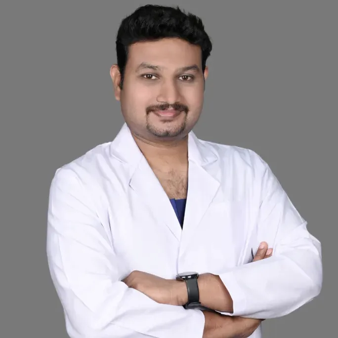
Dr. Pradeep S.
MDS (OMFS) · FHNS · FIBCSOMS
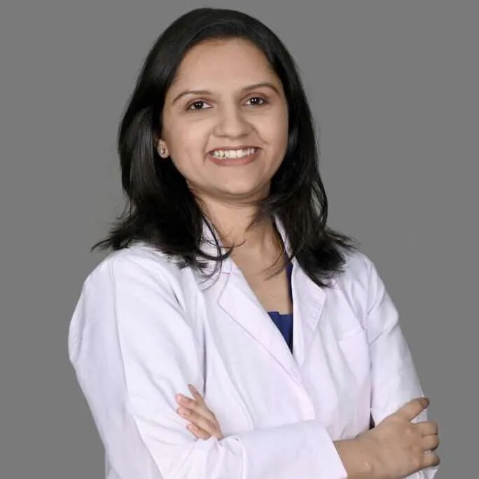
Dr. Kalpa Pandya
MDS (OMFS) · FHNS — Head & Neck Oncology
Authored by
Medically reviewed by