Jaw Bone Cancer: Symptoms, Causes & How It Is Treated

Need expert consultation? Book an appointment with Dr. Pradeep S. or Dr. Kalpa Pandya.
Book AppointmentWhen a patient notices a hard, slow-growing lump on their jawline or experiences unexplained tooth mobility, it often triggers deep anxiety. The jaw is a highly complex anatomical region composed of bone, teeth, nerves, salivary glands, and mucosal linings. Tumours arising in this area can be benign (non-cancerous) yet locally destructive, or they can be malignant (cancerous), requiring immediate, highly coordinated intervention.
Understanding jaw bone cancer is crucial for early detection, which dramatically improves treatment outcomes and preserves facial function. At Mouth Cancer Surgeons in Chennai, Dr. Pradeep S. and Dr. Kalpa Pandya provide comprehensive, dual-surgeon care for patients facing complex jaw pathologies. Consulting at Apollo Main Hospital, Greams Road, Chennai, they manage every stage of the patient's care, from the initial diagnostic biopsy to advanced microvascular reconstruction and dental rehabilitation.
Understanding Jaw Bone Cancer: Malignant Growths vs Benign Jaw Tumours
This guide focuses on malignant disease of the jawbone. If you have been told you have a benign jaw tumour or cyst, such as an ameloblastoma or an odontogenic keratocyst, start with our jaw tumour and cyst specialty page and the ameloblastoma symptoms guide; those conditions are treated differently and carry a very different outlook.
The jawbones—consisting of the mandible (lower jaw) and the maxilla (upper jaw)—are unique because they contain teeth and the specialized tissues responsible for tooth development (odontogenic tissues). Because of this complex cellular environment, the jaw can develop a wider variety of tumours and cysts than any other bone in the human body.
These growths are broadly classified into three categories:
- Benign Odontogenic Tumours and Cysts: These arise from the tissues involved in tooth development. While they are non-cancerous and do not metastasize (spread to other parts of the body), they can be highly aggressive locally, eating away at the surrounding jawbone and shifting teeth out of alignment. Examples include ameloblastomas and odontogenic keratocysts (OKCs).
- Primary Malignant Jaw Bone Cancer: This refers to cancers that originate directly within the bone tissue of the jaw. The most common primary bone cancer is osteosarcoma of the jaw, though chondrosarcomas and Ewing's sarcomas can also occur. These are rare but highly aggressive malignancies.
- Secondary or Metastatic Jaw Cancer: More frequently, jaw bone cancer occurs when a cancer originating in the oral cavity (such as buccal mucosa cheek cancer or floor of mouth cancer) grows deep enough to invade the underlying bone. Alternatively, cancers from distant organs (like the lungs, breasts, or prostate) can metastasize to the jawbone.
Symptoms of Jaw Bone Cancer (and How They Differ From Benign Tumours)
In their early stages, both benign and malignant jaw tumours can be entirely silent, often discovered incidentally during routine dental X-rays. However, as the lesion expands or invades surrounding tissues, several distinct clinical symptoms begin to emerge.
1. Swelling and Facial Asymmetry
The most common presenting symptom is a painless or painful swelling of the jaw, cheek, or the roof of the mouth. The swelling is typically hard to the touch and may cause noticeable changes in facial symmetry.
2. Unexplained Loose Teeth
When a tumour grows within the alveolar bone (the bone that supports the teeth), it destroys the bone structure holding the tooth roots in place. If one or more teeth become loose suddenly, and there is no history of advanced gum (periodontal) disease, this is a major warning sign that requires urgent surgical evaluation.
3. Numbness or Altered Sensation (Paresthesia)
This is a critical clinical symptom. The lower jaw houses the inferior alveolar nerve, which provides sensation to the lower lip and chin. If a tumour or cancer compresses or invades this nerve, the patient will experience numbness, tingling, or a "pins-and-needles" sensation in their lower lip or chin. Unexplained lip numbness is highly suggestive of a malignant jaw lesion.
4. Jaw Pain or Deep Bone Ache
While benign tumours are often painless in their early stages, malignant jaw bone cancers typically cause a persistent, dull, deep-seated ache that does not respond to standard pain relievers or dental treatments like root canals.
5. Difficulty Opening the Mouth (Trismus)
If a tumour involves the muscles of mastication (chewing muscles), the temporomandibular joint (TMJ), or the posterior regions of the jaw, it can severely restrict the patient's ability to open their mouth. This symptom, known as trismus, can make eating, speaking, and oral hygiene highly challenging.
6. Non-Healing Ulcers or Loose Bone
In cases where an oral cancer has invaded the jaw, there may be a visible, painful, non-healing ulcer on the gums overlying the swollen bone. In advanced cases, fragments of dead bone (sequestrum) may become exposed through the gums.
| Symptom / Feature | Benign Jaw Tumours (e.g., Ameloblastoma) | Malignant Jaw Bone Cancer (e.g., Osteosarcoma) |
|---|---|---|
| Growth Rate | Slow, gradual expansion over months or years | Rapid growth, often noticeable week-to-week |
| Pain | Usually painless until it reaches a very large size | Persistent, deep bone pain is common early on |
| Numbness (Lip/Chin) | Very rare | Common due to nerve invasion or compression |
| Tooth Mobility | Teeth shift slowly; roots may show resorption on X-ray | Teeth loosen rapidly; widespread bone destruction |
| Border on X-ray | Well-defined, often with a white border (corticated) | Ill-defined, ragged, "moth-eaten" appearance |
| Metastasis | No risk of spreading to distant organs | Risk of spreading to lungs or regional lymph nodes |
If you or a loved one are experiencing unexplained jaw swelling, loose teeth, or lip numbness, early evaluation is vital. Book an appointment with Dr. Pradeep S. and Dr. Kalpa Pandya at Apollo Main Hospital, Greams Road, Chennai, for an expert diagnostic assessment.
Not sure what your symptom means? Dr. Pradeep S. and Dr. Kalpa Pandya see patients at Apollo Main Hospital, Greams Road, Chennai. Book a consultation or call +91 96633 03747 for an early, unhurried assessment.
Causes and Risk Factors: Why Does Jaw Bone Cancer Develop?
The precise causes of primary jaw bone cancer and benign odontogenic tumours are complex and multifactorial. Unlike many other head and neck cancers, benign jaw tumours are rarely linked directly to lifestyle factors.
- Genetic and Developmental Factors: Many benign jaw tumours and cysts arise from remnants of cells left behind during embryonic tooth development. Errors in cellular signaling during this phase can trigger these dormant cells to begin dividing abnormally, leading to growths like ameloblastomas.
- Prior Radiation Therapy: Patients who have undergone high-dose radiation therapy to the head and neck region for other cancers have a slightly elevated risk of developing secondary primary bone cancers, such as osteosarcoma of the jaw, years later.
- Chronic Irritation and Oral Squamous Cell Carcinoma: For secondary jaw cancer (where oral cancer invades the bone), the primary risk factors are well-established lifestyle habits, particularly tobacco chewing, smoking, and heavy alcohol consumption. These carcinogens damage the mucosal lining of the mouth, leading to oral cancer that eventually erodes into the underlying mandible or maxilla.
- Underlying Bone Disorders: Rare conditions like Paget's disease of the bone or certain genetic syndromes (such as Gorlin-Goltz syndrome) can predispose individuals to developing multiple jaw cysts or osteosarcomas.
How Is Jaw Bone Cancer Diagnosed?
A precise, timely diagnosis is the foundation of successful treatment. Because many jaw lesions share overlapping clinical features, a multi-step diagnostic protocol is essential.
Step 1: Detailed Clinical Examination
At our practice in Chennai, Dr. Pradeep S. and Dr. Kalpa Pandya perform a meticulous extraoral and intraoral examination. They palpate the jaw, check for facial asymmetry, evaluate the mobility of teeth, test nerve sensations along the chin and lip, and check the neck for any enlarged lymph nodes.
Step 2: Advanced 3D Imaging
Standard dental X-rays (like an OPG) are useful for initial screening, but they do not provide the detail needed to plan surgery. We utilize advanced imaging modalities:
- CBCT (Cone Beam Computed Tomography): Provides high-resolution, 3D views of the jawbone, showing the exact relationship of the tumour to the teeth and nerve canals.
- Contrast-Enhanced CT (CECT): Essential for assessing the extent of bone destruction and mapping out the involvement of adjacent soft tissues, muscles, and blood vessels.
- MRI (Magnetic Resonance Imaging): Highly superior for evaluating soft tissue invasion, nerve involvement, and distinguishing fluid-filled cysts from solid bone tumours.
Step 3: Tissue Biopsy (The Gold Standard)
A biopsy is the only definitive way to diagnose jaw bone cancer and bone tumours of the jaw. During this minor procedure, performed under local or general anesthesia, a small sample of the bone or overlying tissue is carefully removed.
An experienced maxillofacial surgeon must perform this biopsy. A poorly planned biopsy incision can spread tumor cells into healthy tissue planes, complicating or compromising the definitive reconstructive surgery that follows.
How Jaw Bone Cancer Is Treated
The primary treatment for almost all jaw tumours—both benign and malignant—is surgical removal. However, the extent of the surgery and the need for additional therapies depend heavily on whether the lesion is benign or malignant.
1. Surgical Management of Benign Jaw Tumours
Benign but aggressive tumours like ameloblastomas require wide surgical clearance. Historically, some surgeons attempted conservative "scraping" (curettage) of these tumours, but this led to exceptionally high recurrence rates.
Today, the standard of care is segmental resection—removing the segment of the jawbone containing the tumour along with a safe margin of healthy bone (typically 1 to 1.5 cm) to ensure no microscopic tumor cells are left behind. This approach minimizes the risk of the tumour returning, but it leaves a defect in the jaw that must be reconstructed.
2. Surgical Management of Malignant Jaw Bone Cancer
Treating malignant jaw bone cancer requires a highly aggressive, multidisciplinary approach:
- Radical Jaw Resection: This involves removing a large portion of the mandible (mandibulectomy) or maxilla (maxillectomy) to ensure completely clear, cancer-free margins.
- Neck Dissection: If the cancer is an oral squamous cell carcinoma that has invaded the bone, or if it is a high-grade sarcoma, the surgeon will also perform a neck dissection to remove lymph nodes in the neck where the cancer is most likely to spread.
- Adjuvant Therapies: Depending on the pathology report, surgery for malignant jaw cancer is often followed by radiation therapy, chemotherapy, or a combination of both (chemoradiation) to destroy any remaining microscopic cancer cells and reduce the risk of recurrence.
Reconstructing the Jaw: Restoring Form, Function, and Quality of Life
Removing a portion of the jawbone can profoundly impact a patient’s life, affecting their appearance, their ability to chew and swallow, and their speech. Therefore, modern maxillofacial oncology does not stop at removing the tumour; it focuses equally on immediate, modern reconstruction.
At Mouth Cancer Surgeons, Dr. Pradeep S. and Dr. Kalpa Pandya specialize in advanced reconstructive and restorative surgery.
Microvascular Free Flap Reconstruction
The gold standard for reconstructing major jaw defects is the fibula free flap. During this highly sophisticated microsurgical procedure:
- A segment of the fibula (the non-weight-bearing bone in the lower leg) is harvested along with its blood vessels (artery and vein).
- The leg bone is precisely shaped and contoured using 3D virtual surgical planning templates to perfectly match the missing segment of the jawbone.
- The shaped bone is secured to the remaining jaw using customized titanium plates.
- Using an operating microscope, the surgeon connects the blood vessels of the leg bone to blood vessels in the neck. This restores immediate blood flow to the newly reconstructed jaw, ensuring the bone survives and heals.
This technique is highly reliable and offers excellent cosmetic and functional outcomes. To understand more about this life-changing procedure, read our detailed guide on fibula free flap jaw reconstruction.
Restoring Your Smile: Dental Implants After Cancer Surgery
Once the reconstructed jawbone has fully healed and fused (typically 4 to 6 months after surgery), patients can undergo dental rehabilitation. Using advanced dental implants and pre-prosthetic surgery, titanium implant posts are placed directly into the newly reconstructed bone. These implants support custom-made dental bridges or dentures, allowing patients to chew, speak, and smile with confidence again. For a deeper look at this phase of recovery, explore our article on dental implants after oral cancer.
Why Choose Mouth Cancer Surgeons in Chennai?
Facing a diagnosis of a jaw tumour or jaw bone cancer can be overwhelming. Choosing the right surgical team is the single most important decision you will make.
Here is why patients across Tamil Nadu and South India trust Dr. Pradeep S. and Dr. Kalpa Pandya:
- The Dual-Surgeon Advantage: At Mouth Cancer Surgeons, you are never cared for by a single doctor or a rotating team of trainees. Both Dr. Pradeep S. and Dr. Kalpa Pandya personally evaluate, plan, and perform your surgery together. This dual-surgeon approach ensures maximum precision, reduced operating times, and highly coordinated care.
- Comprehensive Super-Specialty Expertise: Dr. Pradeep S. is an international board-certified oral and maxillofacial surgeon with over 15 years of dedicated experience in head and neck surgical oncology and microvascular reconstruction. Dr. Kalpa Pandya brings over a decade of specialized experience, having managed over 1000 oral cancer and jaw pathology patients, with a focus on OPMDs, trauma, and dental implant rehabilitation.
- Specialist Infrastructure: Both surgeons consult and operate at Apollo Main Hospital, Greams Road, Chennai (and the Apollo Proton Cancer Centre). This provides patients access to modern intensive care, advanced 3D imaging, digital virtual surgical planning, and a dedicated multidisciplinary oncology board.
- Continuity of Care: From your very first consultation and diagnostic biopsy, through major surgery, microvascular reconstruction, dental implant placement, and years of long-term surveillance, you will see the exact same two surgeons at every single visit.
If you are experiencing symptoms like jaw swelling, loose teeth, or numbness, or if you have received a diagnosis and are seeking a definitive treatment plan or a second opinion, we are here to help.
For personalized treatment options and expert care, consult Dr. Pradeep S. and Dr. Kalpa Pandya — Mouth Cancer Surgeons, Chennai. Call +91 96633 03747 or book an appointment.
References
- Sook-Bin Woo. "Atlas of Oral and Maxillofacial Pathology." Elsevier Health Sciences, 2022.
- National Comprehensive Cancer Network (NCCN). "NCCN Clinical Practice Guidelines in Oncology: Head and Neck Cancers." NCCN Guidelines, 2024. https://www.nccn.org
- Speight, P. M., and Takata, T. "New WHO classification of odontogenic tumours." Paediatric and Developmental Pathology, 2018.
- J. J. G. de Visscher, et al. "Malignant tumors of the jawbones: A long-term follow-up study." Oral Oncology, 2019.
- National Cancer Institute (NCI). "Primary Bone Cancer Treatment - Patient Version." National Institutes of Health, 2023. https://www.cancer.gov
Next step
Concerned about a symptom you read here? Get a specialist opinion in Chennai.
Dr. Pradeep S. and Dr. Kalpa Pandya consult at Apollo Hospitals, Greams Road. Same-day responses on WhatsApp for most enquiries.
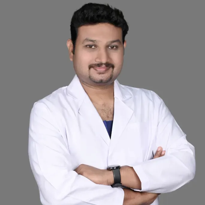
Dr. Pradeep S.
MDS (OMFS) · FHNS · FIBCSOMS

Dr. Kalpa Pandya
MDS (OMFS) · FHNS — Head & Neck Oncology
Authored by
Medically reviewed by