Laser Excision for Oral Precancer: Uses & Recovery
White or red patches in the mouth that do not wipe off and do not heal are called oral potentially malignant disorders, or OPMD. The two best known are leukoplakia (a white patch) and erythroplakia (a red patch). Most of these patches never become cancer, but some do, and there is no reliable way to tell which by looking alone.
When a biopsy shows abnormal cells in a patch, removing it is often recommended. One way to do this is laser excision, most commonly with a CO2 laser. It is a well-established technique for oral precancer, but it is not magic. It does not replace the biopsy that comes first, it does not guarantee the patch will never come back, and it does not remove the need for regular check-ups.
This guide explains when laser excision is used, how it differs from laser ablation, what happens on the day, what healing looks like, and why follow-up continues afterwards.
Leukoplakia and Erythroplakia in Brief
Leukoplakia
Leukoplakia is a white patch on the lining of the mouth that cannot be scraped off and cannot be explained by another condition such as thrush, friction from a sharp tooth or lichen planus. It is closely linked with tobacco smoking and chewing, gutka, betel quid and areca nut. Our patient guide to leukoplakia explains the condition in more detail.
Doctors describe two broad types:
- Homogeneous leukoplakia is flat, thin and evenly white. Its risk of turning into cancer is lower.
- Non-homogeneous leukoplakia is lumpy, wart-like, or speckled with red areas. It carries a higher risk and is watched more closely.
Our article on white patches as a sign of precancer covers the signs and causes.
Erythroplakia
Erythroplakia is a red, smooth or velvety patch that cannot be explained by infection or injury. It is much less common than leukoplakia but more serious. When red patches are biopsied, a large share already show severe dysplasia or early cancer. A red patch in the mouth therefore needs prompt assessment. Read more in our guide to erythroplakia, the red patch in the mouth.
What dysplasia means
The key information comes from the biopsy. The pathologist looks for dysplasia, meaning cells that look abnormal and are not maturing in the normal way. It is graded by how much of the lining is affected:
- Mild dysplasia affects the lower part of the lining.
- Moderate dysplasia extends into the middle part.
- Severe dysplasia or carcinoma in situ affects most or all of the thickness of the lining but has not broken through into the tissue beneath.
In general, the higher the grade, the higher the chance of the patch becoming cancer, and the stronger the case for removing it. Mild dysplasia in a small, flat patch is sometimes watched closely, especially after the person stops tobacco. Moderate or severe dysplasia is usually removed.
Why a Biopsy Always Comes First
Before any laser treatment, the surgeon needs to know what the patch is. A biopsy, taking a small piece of the patch under local anaesthetic, answers several questions:
- Is there dysplasia, and how severe is it?
- Is there already an early cancer?
- Is it something else entirely, such as lichen planus or a fungal infection?
If the biopsy shows an invasive cancer, the treatment is different. Cancer needs a proper cancer operation with wider margins, and sometimes neck surgery, rather than a simple laser removal of the surface.
Large patches can have different grades in different areas, so the surgeon chooses the biopsy site carefully, usually the reddest, thickest or most irregular part. Sometimes more than one biopsy is taken. Our oral cancer biopsy guide explains what to expect.
When Laser Excision Is Recommended
The decision depends on the biopsy result, the appearance of the patch, where it is, its size and your own risk factors. Removal is commonly advised when:
- The biopsy shows moderate or severe dysplasia, or carcinoma in situ.
- The patch is red or speckled, or lumpy and wart-like, even when the first biopsy showed only mild change, because a small biopsy can miss a worse area.
- The patch is at a higher-risk site, such as the side or underside of the tongue or the floor of the mouth.
- The patch persists after a period without tobacco, areca nut and alcohol.
- The patch is changing, growing, thickening or turning red during follow-up.
Laser excision is one of several options. Others include conventional surgery with a scalpel and, for selected low-risk patches, careful monitoring. Your surgeon will explain why one approach suits your situation.
Laser Excision Versus Laser Ablation
These two terms are often confused, but the difference matters.
| Laser excision | Laser ablation (vaporisation) | |
|---|---|---|
| What it does | Cuts the patch out in one piece | Vaporises the surface layer |
| Tissue for the pathologist | Yes, the whole patch | None |
| Can margins be checked? | Yes | No |
| Can hidden early cancer be found? | Yes, if present in the specimen | No, it would be destroyed unseen |
| Typical use | Moderate or severe dysplasia, red or speckled patches, higher-risk sites | Some wide, thin, low-risk patches after biopsy has excluded significant dysplasia |
Because excision gives the pathologist the whole patch to examine, it is usually preferred for higher-risk lesions. Ablation still has a place for some widespread, low-risk patches where cutting out the whole area would be impractical, but only after a biopsy, and with close follow-up afterwards.
How the Procedure Is Done
The CO2 laser produces invisible infrared light that is strongly absorbed by water in the tissue. This lets the surgeon cut very precisely, with the laser sealing small blood vessels as it goes.
Before the procedure
The surgeon examines the patch in good light and marks a margin of normal-looking tissue around it. You will be asked about medicines, especially blood thinners, and given instructions about eating beforehand if sedation or general anaesthetic is planned.
Anaesthetic
- Local anaesthetic is enough for many small patches on the cheek or tongue.
- Sedation or general anaesthetic may be used for larger patches, several patches, or areas that are hard to reach, such as the back of the mouth or the floor of the mouth.
Everyone in the room, including you, wears protective eyewear while the laser is in use.
Removing the patch
The surgeon outlines the margin with the laser, then lifts the patch and cuts beneath it at a controlled depth so that it comes away in one piece. Bleeding is usually minimal because the laser seals small vessels.
Sending the tissue for testing
The specimen is marked so the pathologist knows which edge is which, then sent to the laboratory. The report usually takes about a week.
The wound
Small and medium laser wounds are usually left open to heal on their own rather than being stitched. Healing this way often leaves a soft, flexible scar, which helps keep the tongue and cheek moving normally. Larger wounds may occasionally need other measures, which your surgeon will discuss.
Possible Advantages and Limitations
Advantages often seen with laser excision
- Good control of bleeding, giving the surgeon a clear view of the patch edges.
- A soft scar in many cases, with less tightening than a stitched wound in some sites.
- A specimen for the pathologist, unlike ablation.
- Often done as a day procedure for small patches.
Limitations and risks
- Soreness for one to two weeks.
- Bleeding, usually minor, can occur when the healing coating falls off at around one week.
- Infection is uncommon but possible.
- Scarring or tightness can occur, especially with large wounds.
- Numbness or altered taste is possible near nerves in the tongue or floor of the mouth, and is usually temporary.
- Heat effect at the edges can occasionally make margin assessment slightly harder for the pathologist.
- Recurrence at the same site or new patches elsewhere can still happen.
Recovery Week by Week
Days 1 to 3
- A yellow-white coating forms over the wound. This is normal healing tissue, not pus or infection.
- Expect soreness or a burning feeling, and mild swelling if the tongue or lip was treated.
- Take the painkillers you are prescribed.
- Eat soft, cool, bland food: curd, idli, soft rice and dal, mashed vegetables, cool soups.
- Start gentle salt-water rinses, or the mouthwash you are given, from the day after surgery.
Days 4 to 10
- The coating thins and starts to come away, showing pink tissue underneath.
- Pain usually eases. Speech and eating get easier.
- Move gradually to soft solid foods.
- Keep brushing the rest of your teeth gently, avoiding the wound.
Weeks 2 to 4
- New lining grows across the wound. Small wounds are usually covered by around two to four weeks.
- Most people are back to a normal diet.
- You will have a review appointment to check healing and go through the pathology report.
Do and don't after laser surgery
Do:
- Rinse gently after meals.
- Drink plenty of fluids.
- Take medicines as prescribed.
- Call the team if bleeding does not stop with 15 to 20 minutes of firm pressure using gauze, or if you develop fever, increasing swelling or pain that is getting worse instead of better.
Don't:
- Smoke, chew tobacco, use gutka, pan masala or areca nut, or drink alcohol.
- Pick at or scrub the coating on the wound.
- Have very hot, spicy, sour or crunchy foods in the first week or so.
Have a white or red patch that has not healed in three weeks? Dr. Pradeep S. and Dr. Kalpa Pandya see patients at Apollo Main Hospital, Greams Road, Chennai. Book a consultation or call +91 96633 03747 for an examination and, if needed, a biopsy before any treatment is planned.
Understanding the Pathology Report
At your review, the surgeon goes through the report with you. It confirms:
- The final diagnosis and the highest grade of dysplasia found anywhere in the patch.
- Whether the margins are clear, meaning no abnormal cells at the cut edges.
- Whether any early invasive cancer was found.
If the margins are clear and there is no cancer, you move on to regular follow-up. If dysplasia reaches an edge, the options may include a further small excision or closer monitoring. If an early cancer is found, the surgeon will explain the next steps, which may include wider surgery. Finding this early is one of the main reasons excision is preferred over ablation for higher-risk patches.
Why Follow-Up Continues After Laser Excision
Removing a patch deals with that patch. It does not change the rest of the mouth.
Field change
Years of tobacco, areca nut and alcohol can affect wide areas of the mouth lining, not just the visible patch. Doctors call this field cancerisation, an idea first described in the 1950s. It means that:
- Abnormal cells may remain in normal-looking lining nearby.
- New patches can appear elsewhere in the mouth.
- The treated site itself can develop a patch again.
Recurrence after removal of oral precancer is common enough that regular follow-up is part of standard care, whichever technique was used.
What follow-up involves
Your surgeon will set a schedule based on your biopsy result and risk factors. It is usually more frequent in the first year or two, then gradually spaced out, and for higher-risk patients may continue long term. Each visit includes a careful look and feel of the whole mouth and neck. A new or changing patch may need another biopsy.
What you can do
- Stop tobacco, areca nut and gutka completely. This is the single most important step. Our guide to quitting tobacco after an OPMD diagnosis has practical help, and India's national tobacco quitline is 1800-11-2356.
- Cut down or stop alcohol.
- Check your own mouth once a month in good light, and report any new patch, ulcer or lump that lasts more than three weeks.
- Keep every follow-up appointment, even when everything looks fine.
Key Points
- Leukoplakia and erythroplakia are precancerous patches. Most never become cancer, but some do.
- A biopsy comes first, to grade any dysplasia and rule out cancer.
- Laser excision removes the patch in one piece so the whole of it can be examined. Ablation leaves no specimen and is used only for selected low-risk patches after biopsy.
- Healing usually takes about two to four weeks for small wounds, with a yellow-white coating early on that is normal.
- Laser excision does not guarantee the patch will not return, and it does not prevent cancer elsewhere in the mouth.
- Stopping tobacco and areca nut and keeping regular follow-up remain essential.
You can read more about how precancerous conditions are assessed and treated on our OPMD and oral precancer page.
References
- Warnakulasuriya S, et al. Oral potentially malignant disorders: a consensus report from an international seminar on nomenclature and classification, convened by the WHO Collaborating Centre for Oral Cancer. Oral Diseases. 2021;27(8):1862–1880.
- Mehanna HM, et al. Treatment and follow-up of oral dysplasia: a systematic review and meta-analysis. Head & Neck. 2009;31(12):1600–1609.
- Slaughter DP, Southwick HW, Smejkal W. "Field cancerization" in oral stratified squamous epithelium; clinical implications of multicentric origin. Cancer. 1953;6(5):963–968.
Get a Patch Checked
A patch that is found and assessed early gives you the most options.
Dr. Kalpa Pandya (MDS in Oral & Maxillofacial Surgery, fellowship in head & neck surgical oncology; special interest in OPMD, including leukoplakia, erythroplakia, OSMF and oral lichen planus) and Dr. Pradeep S. (MDS in Oral & Maxillofacial Surgery, FHNS fellowship in head & neck surgical oncology, FIBCSOMS; over 15 years in oral, head and neck cancer surgery) consult at Apollo Main Hospital, Greams Road, Chennai. They examine the mouth, biopsy suspicious patches, explain the report, and plan removal and follow-up where needed.
Book a consultation or call +91 96633 03747.
Next step
Concerned about a symptom you read here? Get a specialist opinion in Chennai.
Dr. Pradeep S. and Dr. Kalpa Pandya consult at Apollo Hospitals, Greams Road. Same-day responses on WhatsApp for most enquiries.
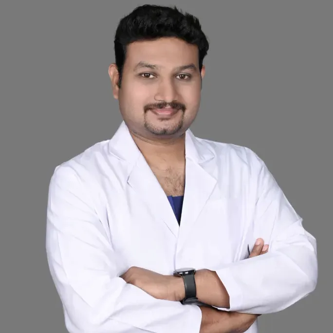
Dr. Pradeep S.
MDS (OMFS) · FHNS · FIBCSOMS
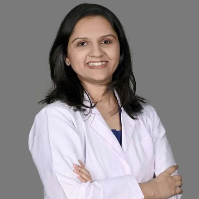
Dr. Kalpa Pandya
MDS (OMFS) · FHNS — Head & Neck Oncology
Authored by
Medically reviewed by