Will I Look Normal After Oral Cancer & Jaw Surgery?

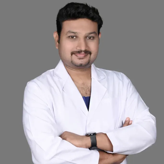
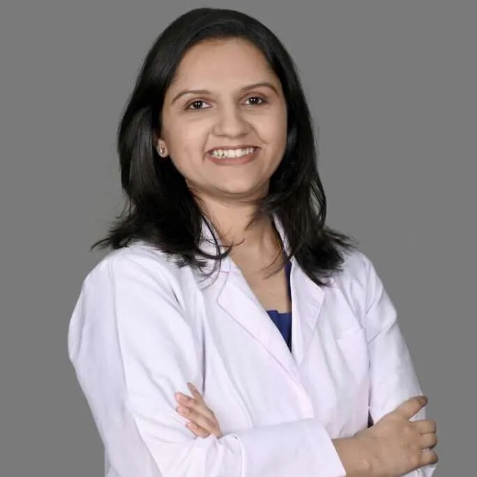
Need expert consultation? Book an appointment with Dr. Pradeep S. or Dr. Kalpa Pandya.
Book AppointmentMany patients tell us that after "will the cancer go?", their next worry is "how will I look when I wake up?" The face is tied to identity and to how we speak, eat and meet people, so this worry is completely natural.
Earlier, oral cancer operations focused mainly on removing the tumour, and sometimes left a visible deficit. Today, in many cases the cancer can be removed and the defect rebuilt in the same operation using the patient's own tissue, with free-flap reconstruction done together with a reconstructive colleague. Some change in appearance may still remain, and your surgeon should tell you honestly what to expect in your case.
What Determines Your Facial Appearance After Surgery?
How your face looks afterwards depends on several things, and reconstruction is planned around your own anatomy.
| Factor | Effect on facial shape |
|---|---|
| Site of the tumour | Cheek and jaw tumours affect the outer contour more than tumours of the floor of mouth or deeper tongue. |
| Amount of bone and tissue removed | Removing a segment of jaw needs bone reconstruction, while a marginal resection keeps the lower border of the jaw. |
| Reconstruction method | Free flaps can match volume well and give stable support, especially when bone is rebuilt; some defects are better handled with other flaps. |
| Radiation | Radiation after surgery can cause skin tightness or volume change over time. |
| Teeth and bone health | Enough bone allows implant-supported teeth, which support the cheek and lip. |
Key Considerations
- Bone Involvement: When a tumor is close to or involves the jawbone (mandible or maxilla), a section of bone must be removed to ensure clear margins. Without reconstruction, removing a segment of jaw can lead to lower-face collapse or asymmetry. Replacing that bone with living bone greatly reduces this. See mandibulectomy for how jaw resection is planned.
- Soft Tissue Volume: Tumors involving the cheek (buccal mucosa), tongue, or soft palate require tissue replacement to maintain natural facial fullness and prevent hollowed contours.
- Incision Placement: Where skin cuts are made determines visibility. Incisions are planned to sit in natural skin creases, expression lines or under the jaw.
Reconstructing the Face: Tissue Transfer Techniques
To restore normal facial shape, surgeons rebuild what was removed using specialized reconstructive methods. For major defects, tissue is transferred from another part of the body—a procedure known as flap reconstruction.
Microvascular Free Flap Reconstruction
Microvascular free flap surgery is widely used for large or complex head and neck defects. Tissue (skin, muscle, or bone) is taken from a donor site with its artery and vein, shaped to fit the defect, and its vessels are joined to vessels in the neck under an operating microscope. It is carried out together with a reconstructive colleague; read more about free flap reconstruction.
Common free flap options include:
- Fibula Free Flap: A portion of the leg bone (fibula) is harvested along with blood vessels and skin. It is contoured to replicate the horseshoe curve of the lower jaw or the complex framework of the upper jaw. Learn more about the fibula free flap jaw reconstruction.
- Anterolateral Thigh (ALT) Flap: Soft tissue from the thigh is used to rebuild large cheek or tongue deficits, providing natural soft-tissue fullness.
- Radial Forearm Free Flap: Thin, pliable skin and tissue from the inner forearm are used to reconstruct delicate oral structures such as the palate or floor of the mouth.
Local and Regional Flaps
For moderate soft tissue defects, regional flaps like the PMMC (Pectoralis Major Myocutaneous) flap or local tissue movement may be used. These are effective, and the choice depends on defect size, your medical history and the goals of reconstruction. Our article on jaw reconstruction: free flap vs PMMC compares them.
See also our reconstructive surgery page.
The Role of 3D Virtual Surgical Planning (VSP)
For selected jaw reconstructions, 3D Virtual Surgical Planning (VSP) and CAD/CAM (Computer-Aided Design and Manufacturing) let the bone reconstruction be planned digitally before surgery. Whether it is useful depends on the case and is discussed during planning.
The usual steps are a high-resolution CT scan, a 3D model of the skull and jaw, virtual removal of the tumour segment with the healthy side mirrored, and then 3D-printed cutting guides and, in some cases, patient-specific plates.
How 3D Planning Preserves Facial Appearance
- Mirroring Healthy Anatomy: If one side of the jaw is affected, the computer program mirrors the healthy opposite side to create a 3D blueprint for reconstruction.
- Custom Cutting Guides: 3D-printed guides are placed on the leg bone (fibula) and jaw during surgery, helping bone cuts match the planned profile.
- Pre-bent Patient-Specific Plates: Titanium plates that hold the reconstructed bone can be shaped to the 3D model to help maintain chin projection and jaw angle.
The aim is that, once swelling settles, the rebuilt jaw supports natural facial contours and lowers the risk of the jaw deviating to one side.
Incisions and Scars: Will They Be Visible?
A common question is whether the cuts will leave visible scars. Surgeons plan incisions along skin tension lines to keep them as inconspicuous as possible, but a scar is never completely absent.
Strategic Incision Placement
- Intraoral Approaches: Whenever clinically appropriate, resections are performed through the inside of the mouth, leaving no external facial scar.
- Submandibular Incisions: Cuts for jaw surgery or neck node removal are placed below the lower border of the jaw, within natural skin creases of the neck. With time these lines are usually hard to notice in everyday conversation.
- Lip-Splitting Approaches: In cases involving extensive tumors of the lower jaw or back of the tongue, a lip-split incision may be necessary. The outline of the lip (vermilion border) is carefully lined up during closure.
Scar Maturation Process
Scars undergo a dynamic healing process over 12 to 18 months:
- Weeks 1–6: Incisions may appear red, elevated, or firm.
- Months 3–6: Softening begins as collagen remodels inside the tissue.
- Months 12–18: Scars flatten, pale in color, and fade into surrounding skin tones.
Post-operative scar care—including topical silicone gels, gentle sun protection, and targeted massage—can help scars settle.
Worried about how you will look after surgery? Dr. Pradeep S. and Dr. Kalpa Pandya see patients at Apollo Main Hospital, Greams Road, Chennai. Book a consultation or call +91 96633 03747 for an assessment and an honest discussion of your reconstruction options.
Restoring Lip Support, Cheeks, and Smile with Dental Rehabilitation
Appearance depends on more than skin and bone. Teeth and the tooth-bearing bone support the lips and cheeks, and when they are missing the cheeks can look sunken and the lips thin.
The Impact of Dental Reconstruction
Rebuilding the jaw gives the foundation, and replacing teeth then restores fullness to the lower face.
- Primary vs. Secondary Dental Implants: Bone flaps such as the fibula allow for placement of osseointegrated dental implants directly into the newly constructed jaw bone. Implants may be placed during the initial tumour surgery or after healing, depending on the case and on whether radiation is planned.
- Fixed Prostheses: Implant-supported bridges can hold teeth securely, helping restore lip and cheek support and a natural smile.
For details on how teeth are restored following oncology procedures, refer to our clinical guide on dental implants after oral cancer or visit the Dental Implants & Pre-Prosthetic Surgery section.
Recovery Timeline: How Your Face Evolves After Surgery
Knowing the usual timeline helps set realistic expectations. Everyone heals at a different pace.
| Phase | What to expect |
|---|---|
| Week 1 to 2 | Marked swelling and dressings; a temporary tracheostomy or feeding tube if needed. The face looks broad. |
| Month 1 | Most swelling settles and incisions heal. Lip movement begins improving. |
| Month 3 to 6 | Soft tissues settle and scars fade from red to pink. Speech and swallowing improve with therapy. |
| Month 12 onwards | Soft-tissue settling is largely complete and scars soften into skin creases. Final dental prosthetics may be in place. |
Stage 1: Immediate Post-Operative (Days 1–14)
Facial swelling is at its peak during the first 72 hours. This is a natural inflammatory response to tissue transfer. Surgical drains and protective dressings may be present around the neck or donor sites. The face may look enlarged or uneven at first; this swelling usually settles steadily.
Stage 2: Early Healing (Weeks 3–8)
As acute edema clears, your natural underlying jaw structure becomes visible. Incisions close completely. Patients begin gentle facial exercises to promote mobility and reduce tissue tightness. For a detailed breakdown of post-surgical timelines, read our guide on oral cancer surgery recovery timeline.
Stage 3: Soft Tissue Settling (Months 3–12)
Microvascular flaps contract slightly and settle naturally into surrounding facial muscular beds over several months. If radiation was part of treatment, mild skin dryness or firmness is managed with moisturisers and physiotherapy exercises.
Stage 4: Long-Term Refinement (Beyond 1 Year)
By about one year, major healing is complete. If you want volume adjustment or scar revision, smaller secondary procedures may be possible.
Psychological Healing and Adapting to Your Appearance
Getting your confidence back is part of recovery, and it takes time. A few things help:
- Set small weekly goals, such as swelling, jaw movement and eating.
- Speak to other patients who have been through surgery; support groups help many families.
- Keep up speech, swallowing and facial exercises with your therapists.
- Raise cosmetic worries openly at follow-up visits, and ask what can be improved later.
Rehabilitation covers speech, swallowing and confidence in daily life. Read more in speech and swallowing therapy after oral surgery.
Planning Cancer Removal and Appearance Together
Reconstruction has to balance removing the disease completely with restoring form and function. Cancer safety comes first; reconstruction is then planned around the defect.
- Dr. Pradeep S. (BDS, MDS, FHNS, FIBCSOMS, PGDCR) is an oral and maxillofacial surgeon with a head and neck surgical oncology fellowship and over 15 years of experience in oral, head and neck cancer surgery.
- Dr. Kalpa Pandya (BDS, MDS, FHNS) is an oral and maxillofacial surgeon with a head and neck surgical oncology fellowship, with interests including oral potentially malignant disorders (OPMD), facial trauma and dental implant rehabilitation.
They plan resection and reconstruction from diagnosis through surgery and follow-up, working with a reconstructive colleague for free flaps and with the wider multidisciplinary team. For life after treatment, see recovery and rehabilitation.
Book a Consultation
A diagnosis of oral cancer or a jaw tumour need not mean giving up your quality of life. With careful planning and reconstruction, many patients return to speaking, eating and social life with an appearance they are comfortable with, even if it is not exactly as before. To discuss your own case, see our oral cancer care page or free flap reconstruction.
- Doctors: Dr. Pradeep S. and Dr. Kalpa Pandya
- Location: Apollo Main Hospital, Greams Road, Chennai
- Phone: +91 96633 03747
- Online booking: Book an Appointment
References
- Hidalgo, D. A. "Fibula Free Flap: A New Method of Mandibular Reconstruction." Plastic and Reconstructive Surgery, vol. 84, no. 1, 1989, pp. 71-79.
- National Comprehensive Cancer Network (NCCN). "NCCN Clinical Practice Guidelines in Oncology: Head and Neck Cancers." NCCN Guidelines
Next step
Concerned about a symptom you read here? Get a specialist opinion in Chennai.
Dr. Pradeep S. and Dr. Kalpa Pandya consult at Apollo Hospitals, Greams Road. Same-day responses on WhatsApp for most enquiries.
Dr. Pradeep S.
MDS (OMFS) · FHNS · FIBCSOMS
Dr. Kalpa Pandya
MDS (OMFS) · FHNS — Head & Neck Oncology
Authored by
Medically reviewed by