A cancerous mouth ulcer typically persists beyond three weeks without healing, is often painless in its early stages, and feels firm or hardened at its base — a quality doctors call induration. Unlike a common canker sore, it may have raised or rolled edges, bleed easily on gentle contact, and will not respond to salt-water rinses, antiseptic gels, or dietary changes. Associated signs such as a new lump in the neck, difficulty moving the tongue, or numbness of the lip add further clinical urgency. Any ulcer that has not healed after three weeks warrants evaluation by a specialist — not continued monitoring at home.
The Worry Behind the Question
Most people who search ‘is my mouth ulcer cancer’ are not being alarmist. They have had a sore in their mouth for two or three weeks — maybe longer — and something about it is not quite right. It has not responded to the usual home remedies. It looks a little different from the ulcers they have had before. And, perhaps most unsettlingly, it does not hurt the way they expected it to.
That kind of worry is rational. It is also the right kind of worry — the sort that prompts people to seek an answer before a problem has time to grow.
Mouth ulcers are among the most common lesions a person can have. The vast majority are completely benign: stress-related canker sores that heal on their own within a week or two, small traumatic injuries from sharp food or a bitten cheek, or minor irritation from a denture that does not fit quite right. In almost every case, these lesions resolve without any treatment at all.
But occasionally, a mouth ulcer does not heal. It persists. It changes, or stays exactly the same while everything around it carries on normally. And in a small but important minority of cases, a non-healing mouth sore is the first visible sign of oral cancer — specifically, oral squamous cell carcinoma, which accounts for the overwhelming majority of mouth cancers diagnosed in India.
The purpose of this article is not to frighten you. It is to give you the specific clinical information you need to make a confident decision about whether to seek an evaluation. There are five features that oral cancer specialists look for when assessing a suspicious ulcer. Knowing them helps you understand your own situation more clearly — and act on it at the right time.
| Feature | Common Mouth Ulcer | Possible Oral Cancer |
| Healing time | 7–14 days | >3 weeks |
| Pain | Usually painful | Often painless early |
| Edges | Smooth | Raised or rolled |
| Base | Soft | Firm or indurated |
| Associated signs | None | Neck lump, numbness |
Who Is at Higher Risk of Oral Cancer?
People with the following factors have a higher risk:
- tobacco smoking
- chewing tobacco or betel nut
- heavy alcohol use
- oral submucous fibrosis
- persistent irritation from sharp teeth or dentures
- HPV infection
5 Warning Signs a Mouth Ulcer May Be More Than a Canker Sore
None of the following features, taken alone, confirms a diagnosis of oral cancer. A diagnosis requires a biopsy. What these signs do is identify which ulcers need that investigation — and which can be reasonably monitored at home for another week or two.
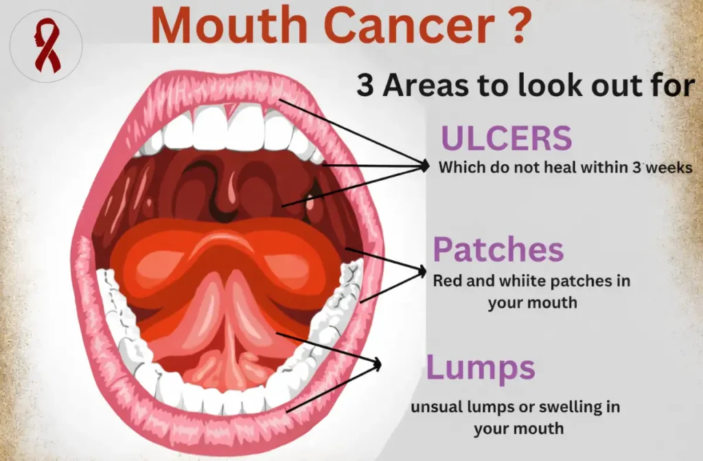
It Has Not Healed in Three Weeks
This is the single most important clinical rule in oral medicine, and it is worth stating plainly: any mouth ulcer that has not healed within 21 days needs a clinical evaluation.
Normal oral mucosa — the moist lining of the mouth — heals quickly. It has an exceptionally rich blood supply, and minor injuries to it repair themselves within one to two weeks in a healthy adult. A canker sore typically peaks around day three or four, begins closing by day seven, and has fully healed by day ten or twelve at the latest. Healing may be slightly slower in someone who is nutritionally depleted, stressed, or immunosuppressed, but it still happens.
An ulcer that simply sits there — unchanged in size and appearance — after three full weeks is not following normal healing biology. Something is preventing repair. That something needs to be identified. One important nuance: if you identified a possible cause — a sharp tooth, an ill-fitting denture — and had it corrected, the ulcer should begin improving within a week of removing the irritant. If it has not, the three-week rule applies regardless.
The Ulcer Is Painless — or Surprisingly Mild
This is the sign that causes the most dangerous delay, because the absence of pain is almost universally interpreted as reassurance. If it does not hurt, people reason, it cannot be serious.
In fact, the reverse is clinically true. A common canker sore is almost always painful — sometimes intensely so. The ulceration of normal oral mucosa exposes nerve endings directly to saliva, food acids, and temperature, which is why a small aphthous ulcer can feel disproportionately uncomfortable relative to its size.
Early-stage oral cancer, by contrast, is frequently painless. The malignant cells infiltrating the submucosal tissue disrupt the local nerve architecture in ways that suppress rather than provoke pain. Many patients with early-stage squamous cell carcinoma describe the lesion as feeling “like nothing” or slightly numb — an odd blankness in an area of the mouth where there should be some sensation. Pain does eventually develop in oral cancer, but it is a feature of more advanced disease — when the tumour has begun invading bone, muscle, or major nerve trunks. By that stage, surgical options are considerably more limited.A painless ulcer that will not heal is not reassuring. It is the pattern that surgeons are most concerned about.
The Ulcer Feels Firm or Hard at Its Base
If you gently press the area around a suspicious ulcer — either directly with a fingertip, or through the cheek wall — a normal benign ulcer will feel soft, consistent with the surrounding tissue. A potentially malignant ulcer often feels noticeably different: firm, almost gritty, as though the tissue beneath has stiffened or thickened.
This quality is called induration. It results from the desmoplastic reaction — a process in which malignant cells stimulate surrounding stromal tissue to produce dense fibrous material, effectively hardening the tissue around and beneath the tumour.
A related finding is fixation: the tissue at the base of the ulcer feels tethered to deeper structures rather than moving freely when gently manipulated. In a benign ulcer, the mucosa slides slightly over the underlying muscle or fat. In a malignant one, that normal slippage is often lost. These features — firmness and fixation — are two of the most reliable indicators that a lesion requires biopsy. They are also entirely detectable during a routine clinical examination, which is precisely why an in-person assessment matters so much more than any photograph or self-examination guide
The Edges Look Raised, Rolled, or Irregular
Look carefully at the border of the ulcer — the junction between the lesion and the surrounding normal tissue. A benign aphthous ulcer has a relatively clean, flat edge that blends into the surrounding mucosa. The border slopes gently toward the ulcerated base.
A potentially malignant ulcer often has a different character at its margin. The edges may appear raised and thickened — rolled inward, almost as though the perimeter of the lesion has been pushed up or everted. This rolled-border appearance is a classic feature of squamous cell carcinoma and reflects the active cellular proliferation occurring at the tumour’s advancing edge.
The surface of the ulcer itself may also look atypical. Granular, irregular, or mixed in colour — areas of white (associated with keratin production), red (indicating thin, fragile mucosa with exposed capillaries), or necrotic tissue at the base. You may also notice that the ulcer bleeds more readily than expected when touched by the tongue or food.
None of these visual features are diagnostic in isolation. But when a raised, irregular border is combined with persistence beyond three weeks and an indurated base, the combination carries a high clinical index of suspicion. A word on surrounding tissue: pay attention to whether there are any white or red patches adjacent to the ulcer. A white patch that cannot be wiped off (leukoplakia) or a velvety red patch (erythroplakia) alongside an ulcer significantly increases concern. Erythroplakia in particular carries a malignant transformation rate that is substantially higher than leukoplakia, and both require specialist assessment.
Other Symptoms Have Appeared Alongside the Ulcer
In early-stage oral cancer, the ulcer itself is often the only finding. But as the disease progresses — and this can happen over weeks to months — additional signs begin to emerge. If any of the following are present alongside a non-healing ulcer, the urgency of evaluation increases significantly.
- A lump or firmness in the neck — typically painless — indicates that cancer cells may have spread to regional lymph nodes. This is one of the most common presentations in patients who arrive at a specialist’s clinic after a period of delay.
- Difficulty moving the tongue or a sensation that the tongue has become restricted suggests involvement of the floor of the mouth or the intrinsic tongue musculature.
- Loosening of a tooth in the vicinity of the ulcer, without any clear dental cause, can indicate periosteal or alveolar bone invasion by an adjacent tumour.
- Numbness of the lower lip, chin, or cheek — sometimes described as a tingling or a ‘deadness’ — can indicate involvement of the inferior alveolar or mental nerve, both of which run through the lower jaw.
- Reduced mouth opening (trismus) may develop if the tumour has infiltrated the pterygoid muscles or the masseteric space.
These are not early-stage findings. Their presence means the evaluation is urgent — not routine.
Common Causes of Mouth Ulcers That Are Not Cancer
The reassuring truth is that the large majority of mouth ulcers have entirely benign explanations. Understanding the common causes helps put the warning signs above into proper perspective.
Traumatic Ulcers
These are the most common mouth ulcers seen in clinical practice. They result from a direct physical injury to the oral mucosa — biting the cheek accidentally, a sharp edge on a broken tooth or a poorly fitting denture, aggressive toothbrushing, or a fragment of hard food. Traumatic ulcers have a clear precipitating cause, appear at the site of injury, and heal within one to two weeks once the cause is removed. If you had the sharp tooth smoothed or stopped wearing the uncomfortable denture and the ulcer resolved within two weeks, it was almost certainly traumatic in origin.
Aphthous Ulcers (Recurrent Canker Sores)
Aphthous stomatitis — the clinical name for recurrent canker sores — affects a significant proportion of the population. These ulcers are characteristically painful, have a clean yellow-white base with a distinct red halo, and appear most commonly on the softer, non-keratinised surfaces of the mouth: the inner lips, the soft palate, and the floor of the mouth. They are often triggered by stress, minor trauma, hormonal changes, or certain foods, and they heal completely without scarring within seven to fourteen days. The key distinguishing feature is the pattern: aphthous ulcers recur in crops, heal, and recur again. A single ulcer that simply does not heal is a different clinical entity entirely.
Nutritional Deficiencies
Deficiencies in iron, vitamin B12, folate, or zinc are well-documented causes of oral ulceration, and they are not uncommon in the Indian population — particularly in vegetarians, women of reproductive age, and those with gastrointestinal malabsorption. Ulcers related to nutritional deficiency tend to be multiple, shallow, and distributed across several mucosal surfaces simultaneously. They typically respond — though sometimes slowly — to appropriate supplementation. If your haematologist or physician has already investigated this pathway and your ulcer has not improved over four weeks of treatment, a different cause needs to be considered.
Viral Infections
The herpes simplex virus is a common cause of oral ulceration, particularly at the time of primary infection. Herpetic ulcers tend to be multiple and clustered, appear on the gingiva or hard palate as well as softer mucosal surfaces, and are often accompanied by fever and systemic malaise during primary infection. Recurrent herpes labialis (cold sores) typically affects the lip border. Hand, foot, and mouth disease — caused by Coxsackievirus — produces small ulcers alongside a characteristic rash and is most common in children. These are self-limiting conditions, and the ulcers resolve as the immune response clears the infection.
When Should You See an Oral Cancer Specialist?
The general guidance is straightforward, and it is worth being direct about it: if your mouth ulcer has not healed within three weeks, book an appointment. Do not wait for a fourth week, or a fifth. The threshold is three weeks, and it exists for a reason.
See a specialist without delay if any of the following apply
- The ulcer has been present for more than three weeks without any sign of healing
- The ulcer is painless, or less painful than you would expect
- A new lump or firmness has appeared in your neck
- The ulcer bleeds easily when touched by your tongue or food
- You have noticed difficulty swallowing, speaking, or opening your mouth fully
- Numbness has developed in the lip, chin, or cheek on the same side
- You use tobacco in any form — cigarettes, beedis, gutka, or pan masala — or drink alcohol regularly
These features do not confirm cancer. They indicate that an evaluation is necessary — and that the evaluation should not be postponed.
In India, tobacco-related oral cancer carries an additional dimension of risk. The habitual chewing of tobacco, areca nut, gutka, and related preparations produces sustained mucosal carcinogen exposure that significantly elevates lifetime oral cancer risk. If you use these products and have a persistent ulcer, the threshold for specialist consultation should be considerably lower than three weeks.
Your first point of contact can be your dentist or oral physician, both of whom are trained to screen for suspicious oral lesions. If they identify any concerning features, they will refer you to an oral and maxillofacial surgeon. You can also book directly with a specialist if you prefer — there is no requirement to wait for a referral when you have clinical concerns that meet the criteria above.
How Is a Suspicious Mouth Ulcer Evaluated?
One of the reasons patients delay seeking evaluation is anxiety about the process itself — the fear that attending a consultation will set in motion a sequence of events they are not prepared for. In reality, the initial assessment is straightforward and causes no significant discomfort.
Clinical Examination
The surgeon will examine all surfaces of the oral cavity under bright light — the lips, cheeks, tongue (including its undersurface and lateral borders), floor of the mouth, hard and soft palate, and gingiva. This takes five to ten minutes and involves no instruments beyond a dental mirror and a light source. The purpose is to identify the location, size, shape, border characteristics, and surface qualities of any lesion.
Palpation
The surgeon will then feel the lesion — and the surrounding tissue — with a gloved finger. This is how induration and fixation are assessed. The neck will also be examined for lymph node enlargement. An enlarged, firm, non-tender lymph node in the submandibular or upper jugular chain is a significant clinical finding that changes management immediately.
Biopsy
If the clinical examination raises concern, a biopsy will be recommended. In most cases this is an incisional biopsy — a small sample of tissue taken from the edge of the lesion under local anaesthesia. The procedure takes ten to fifteen minutes and is performed in the clinic or operating room depending on the site and size of the lesion. The sample is sent to a pathologist, who examines it under a microscope and reports whether malignant cells are present.
A biopsy is not a diagnosis of cancer — it is the means by which a diagnosis is either confirmed or excluded. Many biopsies come back showing benign or pre-malignant changes that can be managed conservatively. The procedure itself is far less frightening than the weeks of uncertainty that precede it.
Imaging
If the biopsy confirms malignancy, the next step is staging — determining the extent and spread of the disease. This typically involves a CT scan of the head, neck, and chest, and sometimes an MRI of the primary tumour site. These investigations guide surgical planning and help determine whether the lymph nodes in the neck need to be addressed at the same time as the primary lesion. For early-stage disease detected promptly, imaging often confirms the reassuring finding that the cancer has remained localised.
Surgeon’s Perspective
In my experience, the pattern I have observed most consistently is not one of sudden dramatic presentations. It is one of gradual delay. A patient notices a sore, waits a week, tries a gel, waits another week. A dentist sees it, prescribes a vitamin supplement, asks the patient to return in a month. By the time the patient reaches my clinic, what began as a lesion smaller than a fingertip has sometimes become a tumour requiring complex reconstructive surgery — a free fibula flap to rebuild the jaw, or an anterolateral thigh flap to restore the tongue.
What I want patients and families to understand is that the difference between Stage I and Stage III oral cancer is not always a matter of years — sometimes it is a matter of months. And the difference in outcome is substantial. A Stage I lesion managed with surgery alone carries a five-year survival rate exceeding eighty percent. By Stage III or IV, when the neck nodes are involved and the tumour has grown into adjacent structures, that figure falls considerably — and the treatment required is far more extensive.
I tell every patient who comes to me with an early lesion the same thing: you did the right thing by coming when you did. The most powerful tool in oral cancer management is not surgery, radiation, or chemotherapy — it is timeliness.— Dr. Pradeep. S., MDS | Oral & Maxillofacial Surgery | Fellow- Head & Neck Oncology | Apollo Hospitals, Chennai
The Most Important Thing You Can Do Today
Mouth ulcers are common. Oral cancer is not common — but it is not rare either, particularly in populations where tobacco and areca nut use are culturally prevalent. The gap between these two realities is navigated by paying attention to the features that distinguish one from the other: duration, painlessness, texture, border irregularity, and associated symptoms.
If your ulcer ticks none of those boxes — it has been present for less than two weeks, it is painful, it has clean soft edges, and you can identify a likely cause — continue home care and monitor it for another week. It will almost certainly heal on its own.
If it ticks one or more of those boxes, stop monitoring and start acting. Book an appointment. The evaluation is not frightening, the biopsy is not painful, and the certainty of knowing is always better than the uncertainty of waiting.
Early oral cancer — detected at Stage I or Stage II — is among the most treatable solid tumour cancers. Surgery alone, in many cases, provides durable cure. The prognosis at that stage is genuinely good. What changes that prognosis, every time, is delay. Do not let a three-week sore become a three-month wait.
Related Reading on This Website
- Oral Cancer — Symptoms, Causes & Risk Factors
- Oral Cancer Screening & Early Detection
- Oral Cancer Biopsy Explained
- Oral Cancer Treatment in Chennai — Complete Guide
- Why Choose a Maxillofacial Surgeon for Oral Cancer
About the Author
Dr. Pradeep S., MDS
Oral & Maxillofacial Surgeon | Fellow – Head & Neck Oncology
Apollo Hospitals, Chennai
Dr. Pradeep S. is an Oral & Maxillofacial Surgeon practicing at Apollo Hospitals, Chennai, with a special interest in oral cancer surgery and head & neck oncology. He evaluates and treats patients with cancers of the tongue, buccal mucosa, gingivobuccal complex, and other oral cavity sites, as well as precancerous oral conditions.
His clinical work includes oncologic resection of oral tumors, neck dissection for lymph node involvement, and multidisciplinary management of oral cancer in collaboration with oncology, radiation therapy, and reconstructive surgery teams.
Dr. Pradeep is actively involved in oral cancer awareness, early detection initiatives, and patient education, with a focus on promoting early diagnosis and improving treatment outcomes.
Clinical Focus
- Oral cancer diagnosis and surgical management
- Tongue cancer and buccal mucosa cancer
- Neck dissection for oral cancer
- Management of oral potentially malignant disorders
- Early detection and screening of oral cancer
Hospital Affiliation
Apollo Hospitals, Chennai
Medical Review
This article has been medically reviewed for clinical accuracy by Dr. Pradeep S., Oral & Maxillofacial Surgeon at Apollo Hospitals, Chennai.
Last medical review: March 2026
Medical Disclaimer
This article is intended for general patient education and does not constitute medical advice. The information provided is not a substitute for a clinical evaluation by a qualified surgeon or physician. If you have concerns about a mouth ulcer or any oral symptom, please seek an in-person assessment from an appropriately trained clinician.